Thursday, November 20, 2008

Week 4

A single water mite was found at the bottom third of the aquarium. It showed very little movement, staying in the same area the entire time I observed it. Its movement was slow, extending one or a few claws perhaps to rotate or scuttle a bare bit. I had identified it as Hydrachna (Smith and Cook, 1991).


The dinoflagellates Perididium willei have increased in number. I have seen at least six this week since the first week.

Other organisms that have increased are the amoeba Succamoeba lucens, which I have seen more than three of, and Vorticella, which have increased beyond counting, all throughout the aquarium.

Much of the diversity and the activity in my aquarium has decreased since Week 3. I have not seen any of the annelids which seemed to be growing and thriving in previous weeks. A few specimens such as Actinosphaeria, Cyclops, and rotifers have not been as present, or even completely absent.


Citations
Smith, I., Cook, R. (1991). Ecology and Classification of North American Freshwater Invertebrates. San Diego: Academic Press, Inc. p547-549. 911 pgs.

Sunday, November 9, 2008

Week 3

When viewing my microaquarium this week it was about two-thirds full with water, there was little activity and I found no new forms of life. I then filled the aquarium to the top and organisms then became more visible.

I discovered a thin, unsegmented annelid trapped among a plant stem and several surrounding algae and other material. It was flexible throughout its entire body and its movement was quick in bursts. It viciously flailed and struggled to break free, but it had already escaped before I could identify it. No camera equipment was available at the time. Later, while viewing the aquarium under a microscope with an attached camera, I found another specimen in a similar scenario:
One other annelid was discovered and at first I had supposed it was an organism under the order Tricladida (Pennak 1989). No kind of gut or any other distinctive features were visible except for the location of its eyes. This made identification dificult and I began questioning my initial guess and with help from Dr. McFarland I had also considered it as Stenostomum.


The debris at the bottom of the aquarium had dimished since I last observed it, and many organisms in this area seemed to no longer be present, or had a lower than previous presence. This includes Cyclops, Perididium, and Actinosphaeria.
However, diatoms Pinnularia and Rhopalodia gibba had greatly increased in number in the bottom third.
Other organisms that have increased in number throughout the entire aquarium include Succamoeba lucens and Vorticella.


Citations
Pennak, R. W. (1989). Fresh-Water Invertebrates of the United States. 3rd Ed. United States: John Wiley & Sons, Inc. p140. 620 pgs.

Sunday, November 2, 2008

Week 2

I have observed quite a few changes in my MicroAquarium since last week. Many organisms have seemed to have vanished and others have flourished. There have especially been a few new organisms that have slipped under the lens from last week:


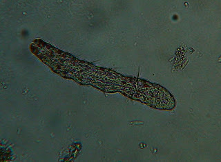
Throughout the top half the aquarium I have seen at least 4 different organisms of the genus Loxophyllum (Patterson 1992). One side is very thick and smooth while the other has several wart-like structures along with the proboscis. Most of these have been seen with smaller organisms inside of them and I have even observed one consuming one of the smaller organisms.


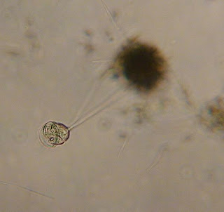
All throughout the aquarium were many single (not colonial) peritrich ciliate protozoa which were assumed as Vorticella by the shape and structure of their bells and springing stalks (Patterson 1992).


Many rotifers have been spotted along the top and middle thirds. These were relatively stationary with only occasional movement in proximal areas only. The organisms were later identified as Rotifer citrinus by the location of their eyes (Ward and Whipple 1918).

A translucent annelid was spotted at the very top of the aquarium at the border of the water. It was very difficult to observe and the only distinctive observation at the time was its red gobules. Another was found behind the glue of the aquarium and again it was very hard to get a clear image of it, but I found many large grouped structures along its body called setae. The next day I came into lab and saw quite a few more, this time in more ideal spots for observation. These annelids are of the genus Aeolosomatidae. Its gut is continuous without distincitve regions and the body is unsegmented (Smith 2001).


There have also been changes in the number of organisms observed the previous week.

The number of Cyclops have decreased, with only two remaining in their likely environment, toward the bottom of the aquarium. There were also a few skeletons in the bottom.

Many more Actinospaerium have sprouted in the aquarium and have moved from only the bottom section upward toward the top the aquarium.

Spirogyra have also reproduced much and have spread up to the middle of the aquarium.


Citations
Patterson, D. J. (1992).Free-Living Freshwater Protozoa. New York: John Wiley & Sons, Inc. p131, 132. Fig. 282, 283. 223 pgs.
Patterson, D. J. (1992). Free-Living Freshwater Protozoa. New York: John Wiley & Sons, Inc. p133. Fig. 233, 235. 223 pgs.
Ward, H. B. (1918). Fresh-Water Biology. Boston: Stabope Press. p618, 619. Fig. 958. 1111 pgs.
Smith, D. G. (2001). Pennak's Freashwater Invertebrates of the United States. New York: John Wiley & Sons, Inc. p275, 276. 638 pgs.